STUB1
STUB1,全稱為STIP1 homology and U-box containing protein 1,也被稱為CHIP,是一種E3泛素連接酶。它在細胞蛋白質穩態中發揮著關鍵作用,通過促進泛素化和降解特定蛋白來調控細胞代謝、分化、增殖、遷移和腫瘤發生發展等多種生理病理過程。STUB1蛋白與多種癌癥類型有關,例如乳腺癌、卵巢癌、胃癌、結直腸癌、肺癌和前列腺癌等。研究表明,STUB1蛋白的表達與癌癥預后存在雙向關聯:在部分癌癥中,高表達STUB1蛋白預示不良預后,因為其會降解抑癌蛋白;而在另一些癌癥中,低表達STUB1蛋白導致預后差,因為其無法降解致癌因子。此外,STUB1蛋白作為治療靶點,可以恢復蛋白穩態并抑制癌癥進展。目前,針對STUB1蛋白的藥物研發尚處于起步階段,但已有研究表明,抑制STUB1蛋白的表達或活性可能有助于提高癌癥免疫療法的療效。
熱銷產品
Recombinant Human E3 ubiquitin-protein ligase CHIP (STUB1) (CSB-EP892480HUe1)
驗證數據
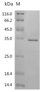
(Tris-Glycine gel) Discontinuous SDS-PAGE (reduced) with 5% enrichment gel and 15% separation gel.
STUB1 Recombinant Monoclonal Antibody (CSB-RA058285A0HU)
驗證數據
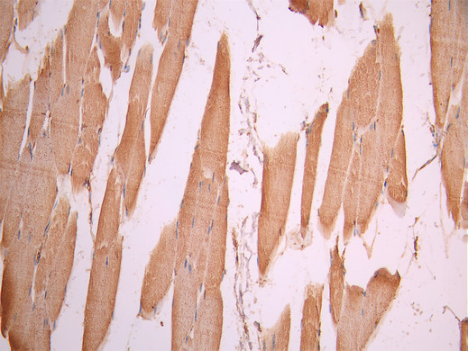
IHC image of CSB-RA058285A0HU diluted at 1:100 and staining in paraffin-embedded human skeletal muscle tissue performed on a Leica BondTM system. After dewaxing and hydration, antigen retrieval was mediated by high pressure in a citrate buffer (pH 6.0). Section was blocked with 10% normal goat serum 30min at RT. Then primary antibody (1% BSA) was incubated at 4°C overnight. The primary is detected by a Goat anti-rabbit polymer IgG labeled by HRP and visualized using 0.05% DAB.
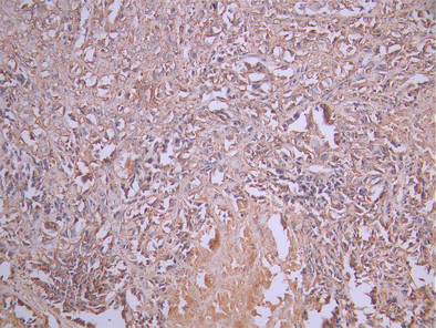
IHC image of CSB-RA058285A0HU diluted at 1:100 and staining in paraffin-embedded human breast cancer performed on a Leica BondTM system. After dewaxing and hydration, antigen retrieval was mediated by high pressure in a citrate buffer (pH 6.0). Section was blocked with 10% normal goat serum 30min at RT. Then primary antibody (1% BSA) was incubated at 4°C overnight. The primary is detected by a Goat anti-rabbit polymer IgG labeled by HRP and visualized using 0.05% DAB.
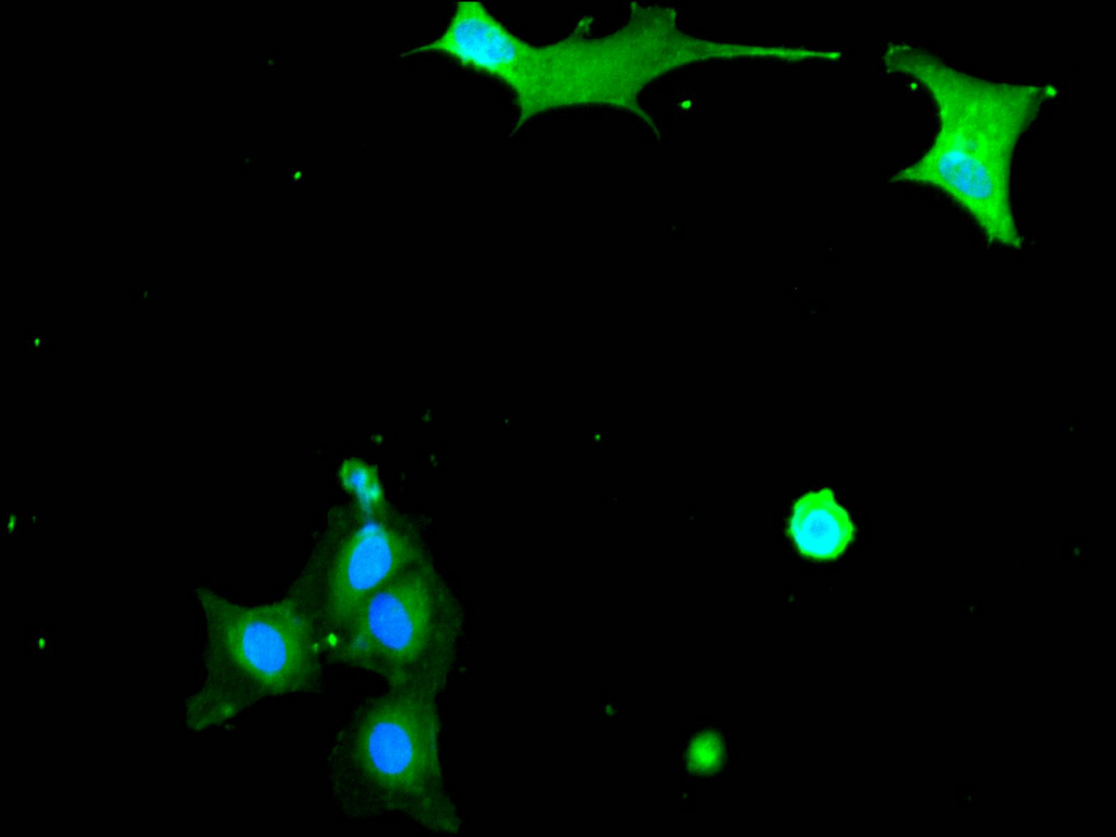
Immunofluorescence staining of MCF-7 cell with CSB-RA058285A0HU at 1:50 , counter-stained with DAPI. The cells were fixed in 4% formaldehyde, permeabilized using 0.2% Triton X-100 and blocked in 10% normal Goat Serum. The cells were then incubated with the antibody overnight at 4°C. The secondary antibody was Alexa Fluor 488-congugated AffiniPure Goat Anti-Rabbit IgG(H+L).
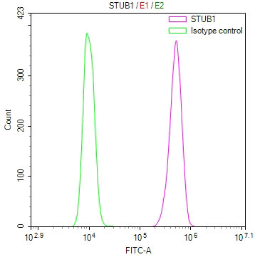
Overlay Peak curve showing Hela cells stained with CSB-RA058285A0HU (red line) at 1:100. The cells were fixed in 4% formaldehyde and permeated by 0.2% TritonX-100 for 10min. Then 10% normal goat serum to block non-specific protein-protein interactions followed by the antibody (1ug/1*106cells) for 45min at 4℃. The secondary antibody used was FITC-conjugated goat anti-rabbit IgG (H+L) at 1/200 dilution for 35min at 4℃.Control antibody (green line) was Rabbit IgG (1ug/1*106cells) used under the same conditions. Acquisition of >10, 000 events was performed.
STUB1 Antibody (CSB-PA892480LA01HU)
驗證數據
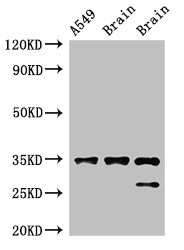
Western Blot
Positive WB detected in: A549 whole cell lysate, Rat brain tissue, Mouse brain tissue
All lanes: STUB1 antibody at 2μg/ml
Secondary
Goat polyclonal to rabbit IgG at 1/50000 dilution
Predicted band size: 35, 28 kDa
Observed band size: 35, 28 kDa
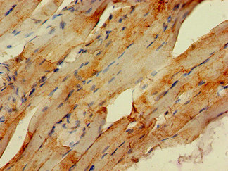
Immunohistochemistry of paraffin-embedded human skeletal muscle tissue using CSB-PA892480LA01HU at dilution of 1:100
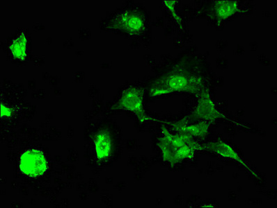
Immunofluorescent analysis of MCF-7 cells using CSB-PA892480LA01HU at dilution of 1:100 and Alexa Fluor 488-congugated AffiniPure Goat Anti-Rabbit IgG(H+L)
STUB1 Antibodies
STUB1 for Homo sapiens (Human)
| 產品貨號 | 產品名稱 | 種屬反應性 | 應用類型 |
|---|---|---|---|
| CSB-PA892480LA01HU | STUB1 Antibody | Human, Mouse, Rat | ELISA, WB, IHC, IF |
| CSB-PA892480LB01HU | STUB1 Antibody, HRP conjugated | Human | ELISA |
| CSB-PA892480LC01HU | STUB1 Antibody, FITC conjugated | Human | |
| CSB-PA892480LD01HU | STUB1 Antibody, Biotin conjugated | Human | ELISA |
| CSB-RA058285A0HU | STUB1 Recombinant Monoclonal Antibody | Human | ELISA, IHC, IF, FC |
| CSB-RA078385A0HU | STUB1 Recombinant Monoclonal Antibody | Human, Mouse, Rat | ELISA, WB, FC, ICC |
STUB1 Proteins
STUB1 Proteins for Gallus gallus (Chicken)
| 產品貨號 | 產品名稱 | 來源 |
|---|---|---|
| CSB-YP719982CH CSB-EP719982CH CSB-BP719982CH CSB-MP719982CH CSB-EP719982CH-B |
Recombinant Chicken STIP1 homology and U box-containing protein 1 (STUB1) | Yeast E.coli Baculovirus Mammalian cell In Vivo Biotinylation in E.coli |
STUB1 Proteins for Homo sapiens (Human)
| 產品貨號 | 產品名稱 | 來源 |
|---|---|---|
| CSB-YP892480HU CSB-BP892480HU CSB-MP892480HU CSB-EP892480HU-B |
Recombinant Human E3 ubiquitin-protein ligase CHIP (STUB1) | Yeast Baculovirus Mammalian cell In Vivo Biotinylation in E.coli |
| CSB-EP892480HUe1 | Recombinant Human E3 ubiquitin-protein ligase CHIP (STUB1) | E.coli |
| CSB-EP892480HU | Recombinant Human E3 ubiquitin-protein ligase CHIP (STUB1) | E.coli |
STUB1 Proteins for Mus musculus (Mouse)
| 產品貨號 | 產品名稱 | 來源 |
|---|---|---|
| CSB-YP895285MO CSB-EP895285MO CSB-BP895285MO CSB-MP895285MO CSB-EP895285MO-B |
Recombinant Mouse STIP1 homology and U box-containing protein 1 (Stub1) | Yeast E.coli Baculovirus Mammalian cell In Vivo Biotinylation in E.coli |