ATP7B
ATP7B(ATPase copper transporting beta),是一種跨膜ATP酶蛋白。它在細胞內負責調節銅元素的轉運和代謝。ATP7B的結構包括多個功能區域,其中包括多個轉運結構域和催化ATP水解的結構域。這些結構域協同作用,使得ATP7B能夠在細胞膜上運輸銅離子。 ATP7B主要表達在肝臟細胞的高銅含量亞細胞器中,如高銅含量的高爾基體和胞質溶酶體。它參與了細胞內銅離子的轉運和分配,將銅從細胞質輸送到高爾基體或從高爾基體釋放到胞質溶酶體。 ATP7B的功能對于維持體內銅平衡和避免銅積累至毒性水平非常重要。銅是生命過程中的關鍵元素,但在高濃度下會對細胞和組織產生毒性作用。因此,ATP7B的異常功能或突變可能導致Wilsons病,這是一種遺傳性銅代謝紊亂疾病。對ATP7B的研究對于理解銅代謝的調控機制以及Wilsons病的發病機理非常重要。同時,ATP7B也成為了治療Wilsons病的一個潛在靶點,研發針對ATP7B的治療方法有望改善這種疾病的治療效果。
熱銷產品
● ATP7B Recombinant Monoclonal Antibody CSB-RA175460A0HU
驗證數據
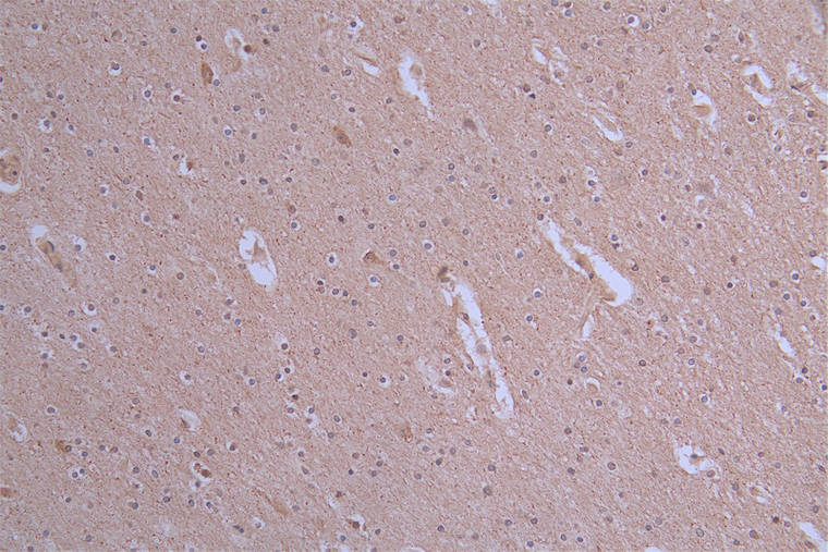
IHC image of CSB-RA175460A0HU diluted at 1:50 and staining in paraffin-embedded human brain tissue performed on a Leica BondTM system. After dewaxing and hydration, antigen retrieval was mediated by high pressure in a citrate buffer (pH 6.0). Section was blocked with 10% normal goat serum 30min at RT. Then primary antibody (1% BSA) was incubated at 4°C overnight. The primary is detected by a Goat anti-rabbit polymer IgG labeled by HRP and visualized using 0.19% DAB
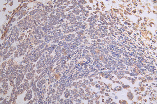
IHC image of CSB-RA175460A0HU diluted at 1:50 and staining in paraffin-embedded human ovarian cancer performed on a Leica BondTM system. After dewaxing and hydration, antigen retrieval was mediated by high pressure in a citrate buffer (pH 6.0). Section was blocked with 10% normal goat serum 30min at RT. Then primary antibody (1% BSA) was incubated at 4°C overnight. The primary is detected by a Goat anti-rabbit polymer IgG labeled by HRP and visualized using 0.19% DAB
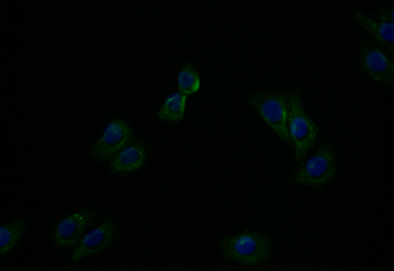
Immunofluorescence staining of HepG2 with CSB-RA175460A0HU at 1:20, counter-stained with DAPI. The cells were fixed in 4% formaldehyde and blocked in 10% normal Goat Serum. The cells were then incubated with the antibody overnight at 4°C. The secondary antibody was Alexa Fluor 494-congugated AffiniPure Goat Anti-Rabbit IgG(H+L).
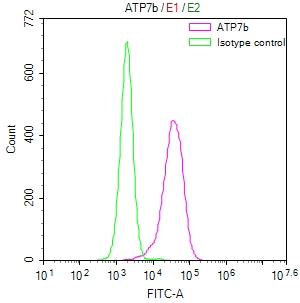
Overlay Peak curve showing HepG2 cells stained with CSB-RA175460A0HU (red line) at 1:50. The cells were fixed in 4% formaldehyde and permeated by 0.2% TritonX-100. Then 10% normal goat serum to block non-specific protein-protein interactions followed by the antibody (1μg/1*106cells) for 45min at 4℃. The secondary antibody used was FITC-conjugated Goat Anti-rabbit IgG(H+L) at 1:200 dilution for 35min at 4℃.Control antibody (green line) was rabbit IgG (1μg/1*106cells) used under the same conditions. Acquisition of >10,000 events was performed.
ATP7B Antibodies
ATP7B for Homo sapiens (Human)
| 產品貨號 | 產品名稱 | 種屬反應性 | 應用類型 |
|---|---|---|---|
| CSB-PA002415ESR2HU | ATP7B Antibody | Human | ELISA, IHC |
| CSB-PA030103 | ATP7B Antibody | Human,Mouse,Rat | IHC, IF, ELISA |
| CSB-RA175460A0HU | ATP7B Recombinant Monoclonal Antibody | Human | ELISA, IHC, IF, FC |
| CSB-RA002415MA1HU | ATP7B Recombinant Monoclonal Antibody | Human | ELISA, IF, FC |
ATP7B Proteins
ATP7B Proteins for Homo sapiens (Human)
| 產品貨號 | 產品名稱 | 來源 |
|---|---|---|
| CSB-YP002415HU CSB-BP002415HU CSB-MP002415HU CSB-EP002415HU-B |
Recombinant Human Copper-transporting ATPase 2 (ATP7B), partial | Yeast Baculovirus Mammalian cell In Vivo Biotinylation in E.coli |
| CSB-EP002415HU | Recombinant Human Copper-transporting ATPase 2 (ATP7B), partial | E.coli |
ATP7B Proteins for Rattus norvegicus (Rat)
| 產品貨號 | 產品名稱 | 來源 |
|---|---|---|
| CSB-YP714496RA CSB-EP714496RA CSB-BP714496RA CSB-MP714496RA CSB-EP714496RA-B |
Recombinant Rat Copper-transporting ATPase 2 (Atp7b), partial | Yeast E.coli Baculovirus Mammalian cell In Vivo Biotinylation in E.coli |
ATP7B Proteins for Mus musculus (Mouse)
| 產品貨號 | 產品名稱 | 來源 |
|---|---|---|
| CSB-YP723743MO CSB-EP723743MO CSB-BP723743MO CSB-MP723743MO CSB-EP723743MO-B |
Recombinant Mouse Copper-transporting ATPase 2 (Atp7b), partial | Yeast E.coli Baculovirus Mammalian cell In Vivo Biotinylation in E.coli |
ATP7B Proteins for Ovis aries (Sheep)
| 產品貨號 | 產品名稱 | 來源 |
|---|---|---|
| CSB-YP896432SH CSB-EP896432SH CSB-BP896432SH CSB-MP896432SH CSB-EP896432SH-B |
Recombinant Sheep Copper-transporting ATPase 2 (ATP7B), partial | Yeast E.coli Baculovirus Mammalian cell In Vivo Biotinylation in E.coli |