PLS1 Antibody
-
中文名稱:PLS1兔多克隆抗體
-
貨號:CSB-PA623928LA01HU
-
規格:¥440
-
圖片:
-
 Western Blot
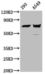
Positive WB detected in: 293 whole cell lysate, A549 whole cell lysate
All lanes: PLS1 antibody at 5.2µg/ml
Secondary
Goat polyclonal to rabbit IgG at 1/50000 dilution
Predicted band size: 71 kDa
Observed band size: 71 kDa -
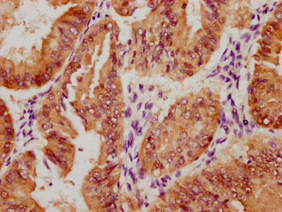
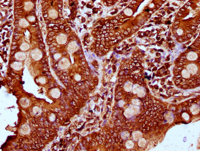 IHC image of CSB-PA623928LA01HU diluted at 1:400 and staining in paraffin-embedded human small intestine tissue performed on a Leica BondTM system. After dewaxing and hydration, antigen retrieval was mediated by high pressure in a citrate buffer (pH 6.0). Section was blocked with 10% normal goat serum 30min at RT. Then primary antibody (1% BSA) was incubated at 4°C overnight. The primary is detected by a biotinylated secondary antibody and visualized using an HRP conjugated SP system.
 -
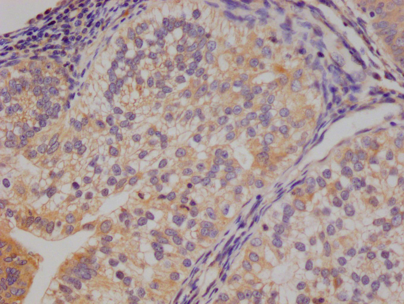
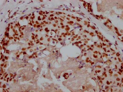
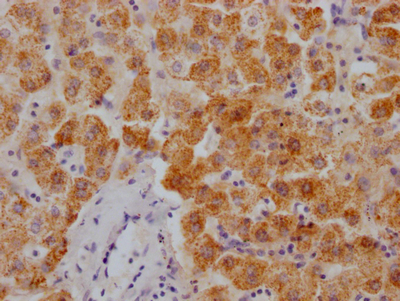
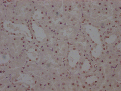
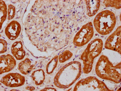 IHC image of CSB-PA623928LA01HU diluted at 1:400 and staining in paraffin-embedded human kidney tissue performed on a Leica BondTM system. After dewaxing and hydration, antigen retrieval was mediated by high pressure in a citrate buffer (pH 6.0). Section was blocked with 10% normal goat serum 30min at RT. Then primary antibody (1% BSA) was incubated at 4°C overnight. The primary is detected by a biotinylated secondary antibody and visualized using an HRP conjugated SP system.
 -
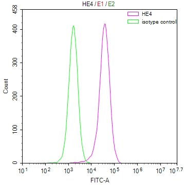
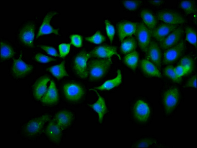 Immunofluorescence staining of A549 cells with CSB-PA623928LA01HU at 1:133, counter-stained with DAPI. The cells were fixed in 4% formaldehyde, permeabilized using 0.2% Triton X-100 and blocked in 10% normal Goat Serum. The cells were then incubated with the antibody overnight at 4°C. The secondary antibody was Alexa Fluor 488-congugated AffiniPure Goat Anti-Rabbit IgG(H+L).
 -
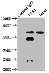 Immunoprecipitating PLS1 in 293 whole cell lysate
Lane 1: Rabbit control IgG instead of CSB-PA623928LA01HU in 293 whole cell lysate. For western blotting, a HRP-conjugated Protein G antibody was used as the secondary antibody (1/2000)
Lane 2: CSB-PA623928LA01HU (6µg) + 293 whole cell lysate (500µg)
Lane 3: 293 whole cell lysate (20µg)
-
-
其他:
產品詳情
-
產品名稱:Rabbit anti-Homo sapiens (Human) PLS1 Polyclonal antibody
-
Uniprot No.:
-
基因名:PLS1
-
別名:PLS1 antibody; Plastin-1 antibody; Intestine-specific plastin antibody; I-plastin antibody
-
宿主:Rabbit
-
反應種屬:Human
-
免疫原:Recombinant Human Plastin-1 protein (1-143AA)
-
免疫原種屬:Homo sapiens (Human)
-
標記方式:Non-conjugated
本頁面中的產品,PLS1 Antibody (CSB-PA623928LA01HU),的標記方式是Non-conjugated。對于PLS1 Antibody,我們還提供其他標記。見下表:
-
克隆類型:Polyclonal
-
抗體亞型:IgG
-
純化方式:>95%, Protein G purified
-
濃度:It differs from different batches. Please contact us to confirm it.
-
保存緩沖液:Preservative: 0.03% Proclin 300
Constituents: 50% Glycerol, 0.01M PBS, pH 7.4 -
產品提供形式:Liquid
-
應用范圍:ELISA, WB, IHC, IF, IP
-
推薦稀釋比:
Application Recommended Dilution WB 1:500-1:5000 IHC 1:200-1:500 IF 1:50-1:200 IP 1:200-1:2000 -
Protocols:
-
儲存條件:Upon receipt, store at -20°C or -80°C. Avoid repeated freeze.
-
貨期:Basically, we can dispatch the products out in 1-3 working days after receiving your orders. Delivery time maybe differs from different purchasing way or location, please kindly consult your local distributors for specific delivery time.
-
用途:For Research Use Only. Not for use in diagnostic or therapeutic procedures.
產品評價

相關產品
靶點詳情
-
功能:Actin-bundling protein. In the inner ear, it is required for stereocilia formation. Mediates liquid packing of actin filaments that is necessary for stereocilia to grow to their proper dimensions.
-
基因功能參考文獻:
- upregulated in eosinophils from atopic dermatitis patients PMID: 27304220
- In this study, the authors found that the actin filament bundling abilities of PLS1 and PLS2 were similarly sensitive to Ca(2+) (pCa50 ~6.4), whereas PLS3 was less sensitive (pCa50 ~5.9). PMID: 28694070
-
亞細胞定位:Cytoplasm. Cell projection, stereocilium.
-
組織特異性:In small intestine, colon, and kidney; relatively lower levels of expression are detected in the lung and stomach.
-
數據庫鏈接:
Most popular with customers
-
-
YWHAB Recombinant Monoclonal Antibody
Applications: ELISA, WB, IHC, IF, FC
Species Reactivity: Human, Mouse, Rat
-
Phospho-YAP1 (S127) Recombinant Monoclonal Antibody
Applications: ELISA, WB, IHC
Species Reactivity: Human
-
-
-
-
-